
Youngsun Kong, Ph.D.
Postdoctoral Research Fellow, Biomedical Engineering
University of Connecticut, Storrs, CT
From Physiological Signals to Objective Human State Assessment
Email · CV · Google Scholar · GitHub · LinkedIn
(18 first or co-first author)
as PI or Co-I
About
I am a biomedical engineer with a foundation in computer science. During my Ph.D. in Biomedical Engineering at the University of Connecticut, I brought signal processing and machine learning to a clinical problem that still lacks reliable objective measures: pain. My doctoral work used physiological signals to study dental pain and pulp status, and this research has since expanded into an NIH F32-supported program. In parallel, in collaboration with the U.S. Navy, I apply the same approaches to human performance under physiological and environmental stress. Across these areas, one question drives my research: how can we turn complex signals from the body into objective measurements we can trust?
Research
I develop computational frameworks that combine signal processing, machine learning, and AI to extract reliable and physiologically meaningful information from complex biosignals. I also design and conduct controlled human experiments to characterize physiological responses and validate these methods. My research spans robust physiological sensing, quantitative signal characterization, objective pain and nociception assessment, and human-state and performance monitoring under physiological and environmental stress. My long-term goal is to develop physiological assessments that account for individual differences and remain reliable across clinical and human-performance settings.
Physiological Signal Characterization
I develop methods to extract sensitive and interpretable information from physiological recordings. This work examines how skin conductance and skin nerve activity change during controlled stimulation, turning raw signals into quantitative measures of autonomic and pain-related responses.
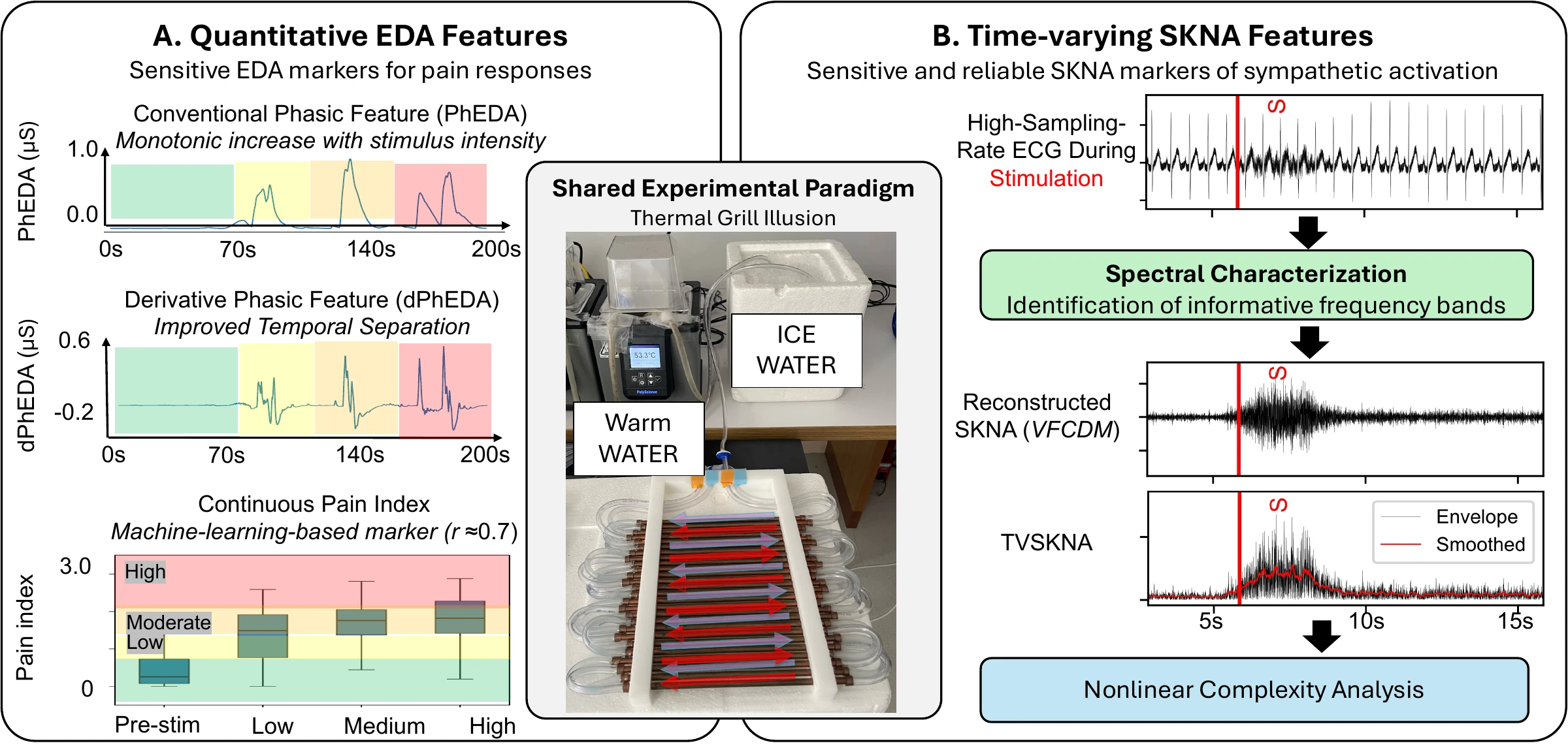
- Sensitive physiological indices of pain based on differential characteristics of electrodermal activity IEEE TBME, 2021 Co-first author
- Objective pain stimulation intensity and pain sensation assessment using machine learning classification and regression based on electrodermal activity Am J Physiol Regul Integr Comp Physiol, 2021 Co-first author
- A new approach to characterize dynamics of ECG-derived skin nerve activity via time-varying spectral analysis IEEE Trans Affective Computing, 2025 First author
- Complexity analysis of skin nerve activity for quantitative assessment of acute sympathetic nervous system activation Sensors, 2026 First author
- PI-AMFM: Permutation-invariant learning for variable-cardinality AM-FM mode decomposition in biomedical signal analysis arXiv preprint, 2026 First author
Translational Pain Assessment
I use physiological signals to develop objective measures of pain-related responses during dental testing. This work progresses from distinguishing dental stimulation from stress using electrodermal activity (EDA) to combining multiple physiological signals and individual characteristics for more comprehensive pain assessment.
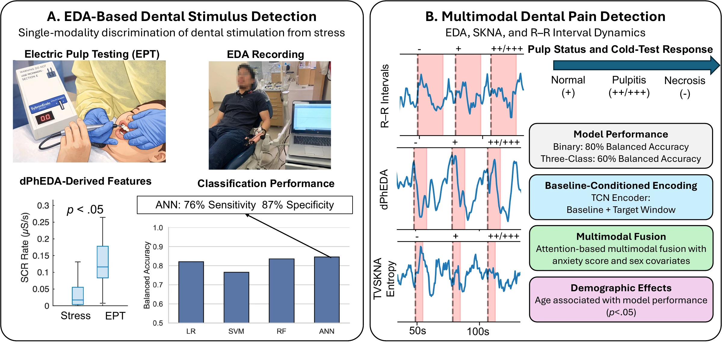
- Differentiating between stress- and EPT-induced electrodermal activity during dental examination Comput Biol Med, 2023 First author
- A multimodal autonomic sensing framework for objective assessment of patient responses to dental pulp stimulation arXiv preprint, 2026 (under review) First author
Robust Physiological Sensing
I develop methods that help physiological measurements remain useful when recordings contain movement-related interference or other noise. These approaches either suppress contamination to recover useful information or identify unreliable intervals before further analysis.
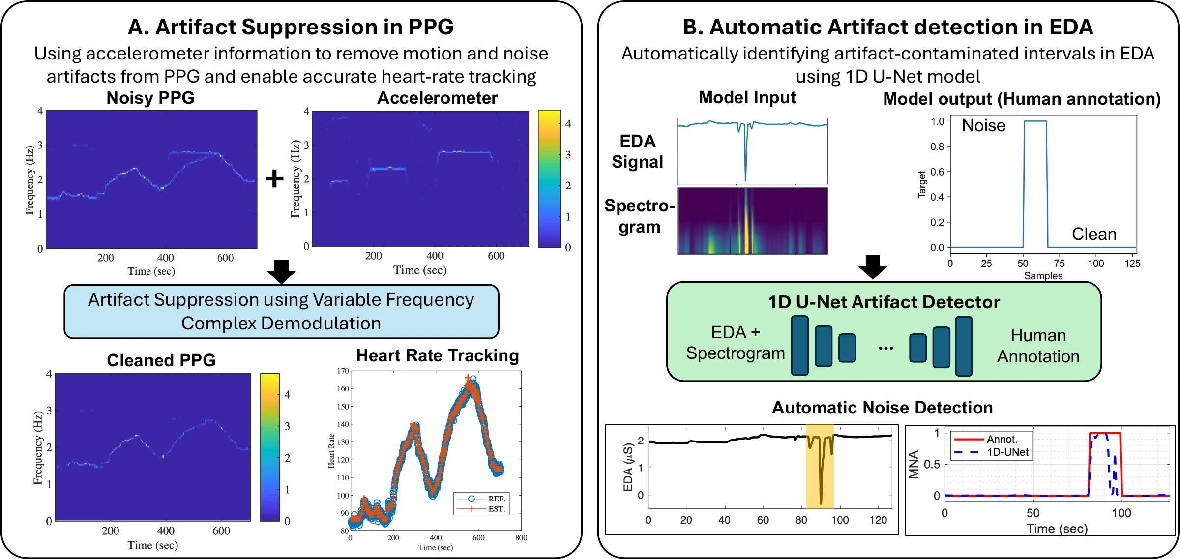
- Heart rate tracking using a wearable photoplethysmographic sensor during treadmill exercise IEEE Access, 2019 First author
- Automatic motion artifact detection in electrodermal activity signals using 1D U-net architecture Comput Biol Med, 2024 First author
Human Nociception and Pain Dynamics
I use controlled human experiments to examine how perceived pain and autonomic responses evolve during sustained stimulation. Related machine-learning work tests whether these physiological responses can distinguish stimulation conditions designed to preferentially engage different sensory nerve fibers.
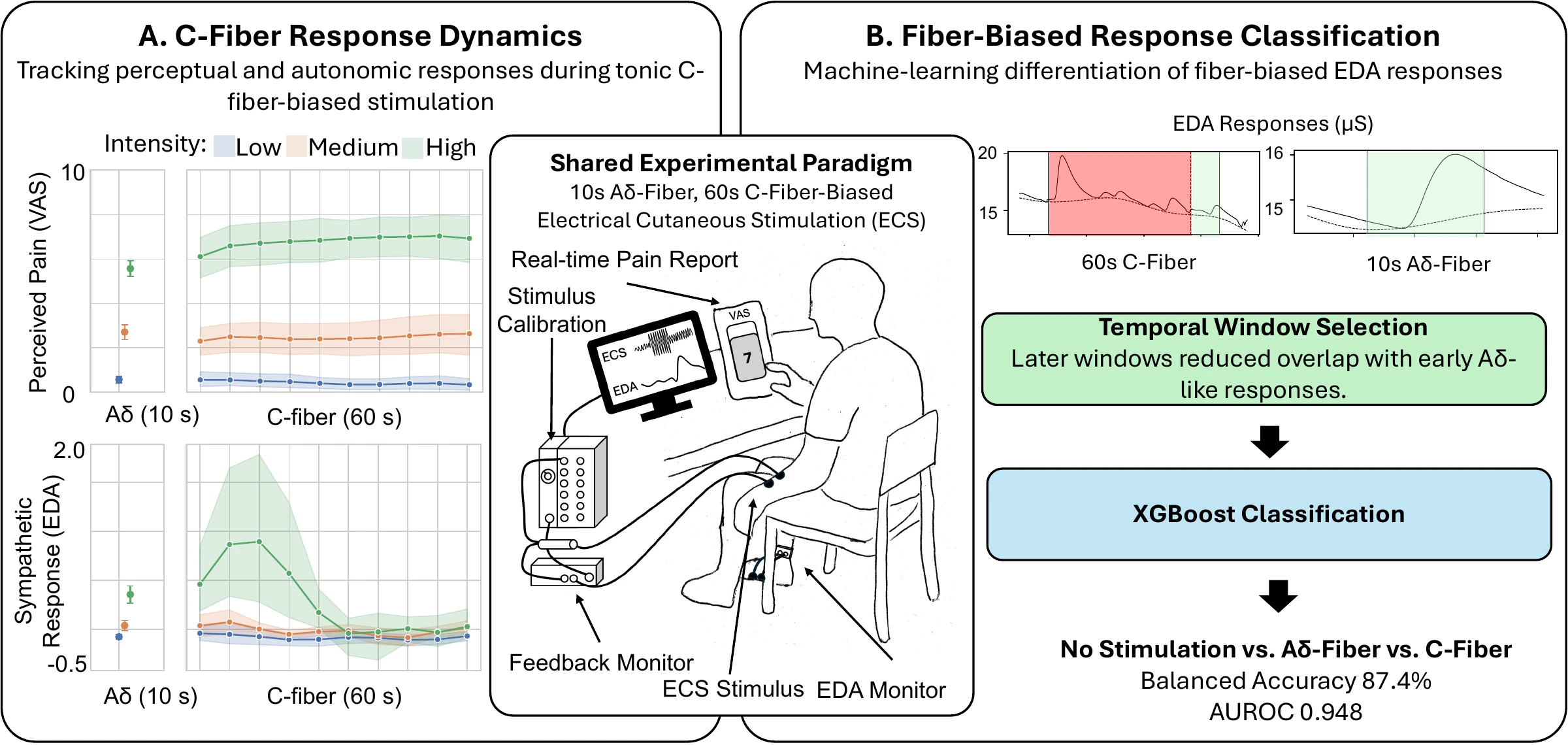
- A tonic electrocutaneous stimulation paradigm for graded sensory C-fiber engagement in healthy subjects Front Pain Res, 2026 Co-first author
- Differentiation of peripheral nerve fiber activation through electrodermal activity in healthy adults Biocybern Biomed Eng, 2026 Co-first author
Human Performance Under Challenging Conditions
I investigate how prolonged wakefulness and cold exposure affect human performance, using behavioral measurements, physiological signals, and machine learning. Collaborative engineering projects extend this work toward wearable monitoring and early warning of physiological risk in extreme environments. This line of work has been conducted in collaboration with U.S. Navy researchers.
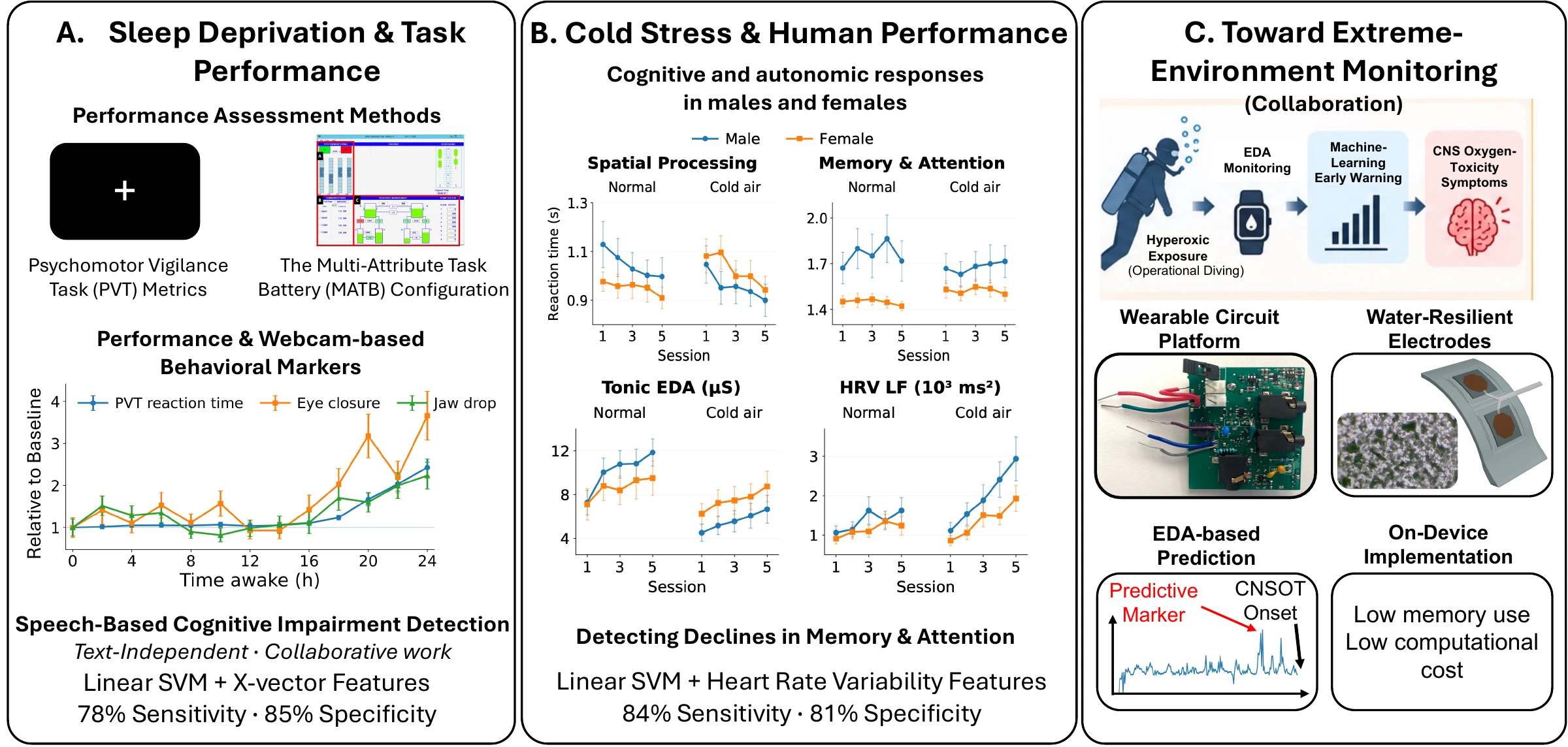
- Facial features and head movements obtained with a webcam correlate with performance deterioration during prolonged wakefulness Atten Percept Psychophys, 2021 First author
- Multi-Attribute Task Battery configuration to effectively assess pilot performance deterioration during prolonged wakefulness Inform Med Unlocked, 2022 First author
- Sex differences in autonomic functions and cognitive performance during cold-air exposure and cold-water partial immersion Front Physiol, 2024 First author
- Detection of cognitive performance deterioration due to cold-air exposure in females using wearable electrodermal activity and electrocardiogram Biosensors, 2025 First author
- Acted sleepy speech vs. real sleepy speech: Human perception and machine prediction for sleepiness estimation Int J Hum-Comput Stud, 2026 Co-author
- Design and validation of a multimodal wearable device for simultaneous collection of electrocardiogram, electromyogram, and electrodermal activity Sensors, 2022 Co-author
- Prediction of central nervous system oxygen toxicity symptoms using electrodermal activity and machine learning Biocybern Biomed Eng, 2024 Co-author
- Mechanically robust superhydrophobic coatings via dual-step deposition for electrodermal activity (EDA) electrodes immersible in saltwater Materials & Design, 2026 Co-author
Funding & Awards
Awarded Funding
- NIH/NIDCR Ruth L. Kirschstein Postdoctoral Individual NRSA (F32), Role: PI 2024–2027
- Defense Health Agency STTR Program, Phase II, Role: Co-I 2024–2026
Pending / Submitted
- NIH/NIDCR K99/R00 Pathway to Independence Award (submitted), Role: PI
- ONR/NIUVT (submitted), Role: Co-PI
Selected Awards
- Top 10 Finalist, Young Professional Paper Competition, IEEE EMBC 2025
- NIH Ruth L. Kirschstein Postdoctoral Individual NRSA (F32) 2024
- Best Paper Award, 3rd Place, IEEE-EMBS International Conference on Body Sensor Networks 2023
Publications
44 peer-reviewed journal articles (18 first/co-first; 10 corresponding). Key papers are listed under each research theme above; the full list is in my CV and on Google Scholar.
Featured Review
- Kong, Y., & Chon, K. H. (2024). Electrodermal activity in pain assessment and its clinical applications. Applied Physics Reviews, 11(3), 031316.
Background
Appointments
- Postdoctoral Research Fellow (NIH NRSA F32), University of Connecticut 2024–Present
- Postdoctoral Research Associate, University of Connecticut 2022–2024
Education
- Ph.D., Biomedical Engineering — University of Connecticut 2022
- B.S. & M.S., Computer Science and Engineering — Soonchunhyang University 2015/2017
Teaching & Service
Teaching
-
Guest Lecturer, University of Connecticut
BME 6086, Advanced Biomedical Signal Processing · ENGR 1166, Foundations of AI: Machine Learning 2022–Present
Mentoring
-
Mentored 10+ undergraduate and graduate researchers, leading to co-authored publications and presentations
Highlight: a mentee’s IEEE BSN Best Paper Award (3rd place) -
Senior Design Co-Mentor, UConn Biomedical Engineering
Two-student team developing a smartwatch-based pain monitoring system 2025–2026
Editorial & Review Service
- Associate Editor, Frontiers in Pain Research
- Associate Editor, IEEE Engineering in Medicine and Biology Conference (EMBC)
- Special Issue Editor, Sensors (Advanced Signal Processing for Affective Computing)
- Guest Editor, Frontiers in Pain Research (Sensors and AI for Objective Pain Detection)
- Program Committee Member, AI4Pain Challenge at ICMI 2025 and IEEE ACII 2026
- Ad hoc reviewer: Nature Communications, IEEE Journal of Biomedical and Health Informatics, Scientific Reports, and others
Outreach
- PBS NOVA (Season 50, Episode 9): co-presented a thermal grill illusion demonstration from my pain research, featured in the aired segment
Contact
Department of Biomedical Engineering, University of Connecticut, Storrs, CT